Branchial Cleft Cysts
Disclaimer: Disclaimer: This leaflet provides general information and is intended for educational purposes only. It should not be used as a substitute for professional medical advice, diagnosis, or treatment. Always seek the advice of a qualified healthcare professional for any health concerns or before making any decisions related to your health or treatment.
This leaflet may contain links to external websites or resources (e.g. YouTube) for demonstration purposes; however, these links are provided for information only. Clinicol.co.uk is not affiliated with, does not endorse, and is not responsible for the content, accuracy, or copyright compliance of these external sources. Use of these external links is at your own discretion and risk.
This leaflet is designed to provide information about branchial cleft cysts. It explains what they are, how they are diagnosed, and the treatment options available. This leaflet is a general guide, and your individual circumstances may vary. Always discuss your specific case with your doctor or surgeon (such as an ENT or Head and Neck Surgeon).
Overview
A branchial cleft cyst is a type of lump that develops in the neck. It's a congenital anomaly, which means it's present from birth. These cysts are formed during the very early stages of a baby's development in the womb.
Think of the developing neck and face as being built from a series of arches, a bit like the arches of a bridge. These are called branchial arches. Normally, these arches fuse together seamlessly. However, sometimes small gaps or pockets remain. If the gap forms a closed pocket, it can fill with fluid, creating a branchial cleft cyst. If the gap forms a connection to the skin or inside the throat, it's called a branchial cleft sinus or fistula. This leaflet focuses mainly on cysts, but much of the information also applies to sinuses.
Branchial cleft cysts are almost always benign (non-cancerous). However, they can cause problems if they become infected or grow large enough to press on nearby structures. They can appear at any age, although they are most commonly noticed in children or young adults.
Symptoms and Causes
What causes them?
As mentioned, these cysts are caused by incomplete fusion of the branchial arches during fetal development. There's no known way to prevent them, and they are not caused by anything the mother did or didn't do during pregnancy. It's simply a variation in how the neck develops. In some cases, branchial cleft anomalies can be associated with other conditions, such as branchio-oto-renal (BOR) syndrome, which can affect hearing and kidney development. Your doctor will assess for this if necessary.
What are the symptoms?
The symptoms can vary depending on the location and size of the cyst, and whether it's infected. Here are some common signs:
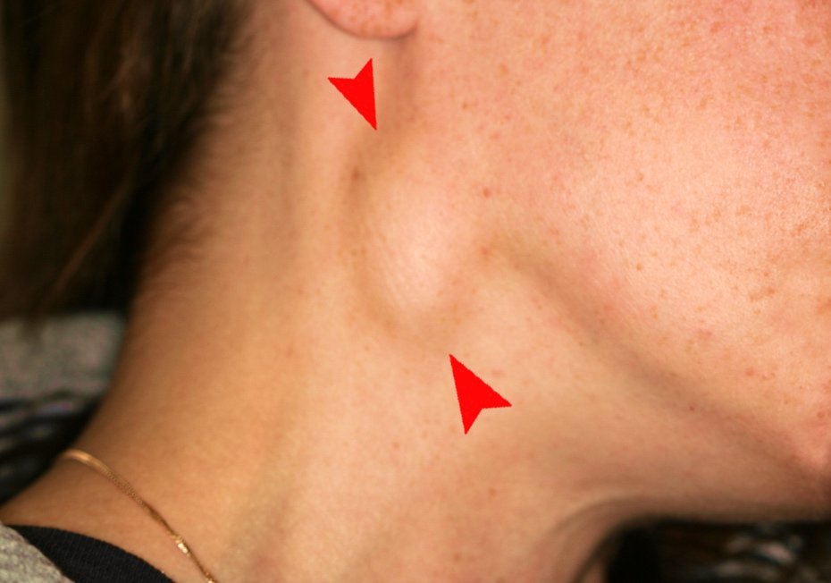
- A painless lump in the neck: This is the most common symptom. The lump may be smooth and round, and it might move slightly when touched or when swallowing. It can be on either side of the neck.
- Swelling: The lump may gradually get bigger over time, or it may suddenly swell if it becomes infected.
- Skin changes: If a sinus tract is present, there may be a small opening on the skin, sometimes with drainage of mucus-like fluid. This is more common with sinuses than cysts.
- Infection: If the cyst becomes infected, it can become:
- Red
- Painful and tender
- Swollen
- Warm to the touch
- May discharge pus
- Difficulty swallowing (dysphagia): A large cyst, especially if infected, can sometimes make swallowing uncomfortable.
- Noisy breathing (stridor): In rare cases, a very large cyst can press on the airway, causing noisy breathing. This is more of a concern in infants.
- Recurrent neck infections: In some patients they may present as recurring neck infections (abscesses).
Types of Branchial Cleft Cysts
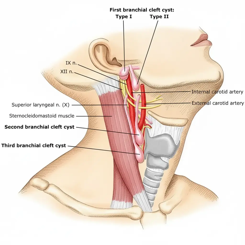
There are different types, classified by which branchial arch they originate from:
- First branchial cleft cysts: These are less common and are usually found near the ear, jawline, or ear canal. They may have a sinus tract opening above or below the jawline.
- Second branchial cleft cysts: These are the most common type. They typically appear on the side of the neck, below the jaw, and in front of a large neck muscle called the sternocleidomastoid.
- Third and Fourth branchial cleft cysts: These are rarer and located lower in the neck, near the thyroid gland. They can sometimes present with recurrent infections in the region of the thyroid.
Diagnosis and Investigations
If you or your child has a lump in the neck, it's important to see a doctor (GP) to get it checked. The doctor will:
- Take a Medical History: They'll ask about the lump, how long it's been there, any other symptoms, and your general health.
- Perform a Physical Examination: They'll examine the lump and the rest of the neck and head area.
Investigations:
To confirm the diagnosis and rule out other conditions, the doctor may recommend some tests:
- Ultrasound Scan: This is often the first test. It uses sound waves to create an image of the cyst and surrounding tissues. It's painless and doesn't involve radiation.
- CT Scan (Computed Tomography Scan): This uses X-rays to create detailed images of the neck. It may be used if the ultrasound is unclear or if the doctor needs more information about the cyst's size and location, especially before surgery. A contrast dye may be injected into a vein to improve the image.
- MRI Scan (Magnetic Resonance Imaging): This uses strong magnets and radio waves to create detailed images. It's particularly good for showing soft tissues and may be used if a CT scan is not suitable or if more detail is needed.
- Fine Needle Aspiration (FNA): In some cases, the doctor may use a thin needle to take a sample of fluid from the cyst. This fluid can be examined under a microscope to help confirm the diagnosis and rule out infection. This is less common for straightforward cysts in children.
- Endoscopy: If a sinus tract is suspected to connect to the inside of the throat, a special examination under anaesthesia, called endoscopy may be needed. This is less commonly required.
- Hearing Test: If a first branchial cleft cyst is suspected, a hearing test may be required.
It's important to remember that not all of these tests will be necessary for every patient. Your doctor will decide which tests are appropriate for your individual situation.
Management and Treatment
The main treatment for branchial cleft cysts and sinuses is surgical removal. However, the approach depends on whether the cyst is infected.
1. If the cyst is NOT infected:
- Observation: Very small, asymptomatic cysts might be monitored without immediate surgery, particularly in very young children. However, there's always a risk of future infection, so surgery is usually recommended.
- Surgery (Excision): The standard treatment is to surgically remove the entire cyst and any associated sinus tract. This is done under general anaesthesia (you'll be asleep). The surgeon will make an incision in the neck, carefully remove the cyst, and close the incision with stitches. The exact location and size of the incision will depend on the cyst's location.
- First branchial cleft cysts: may require removal of part of the parotid gland.
- Third and Fourth branchial cleft cysts: may require removal of part of the thyroid gland.
2. If the cyst IS infected:
- Antibiotics: The first step is to treat the infection with antibiotics. These are usually taken by mouth (orally). Common antibiotics used in the UK include:
- Flucloxacillin: (Prescription-only) Usually taken four times a day. The dose will depend on the patient's age and weight.
- Co-amoxiclav: (Prescription-only) Usually taken three times a day. Again, the dose depends on age and weight.
- Clarithromycin: (Prescription-only) An alternative for patients allergic to penicillin. Usually taken twice a day.
- Erythromycin: (Prescription-only, but some formulations are available over-the-counter for other conditions, not for treating branchial cleft infections). Used as an alternative if allergic to penicillin. Often taken four times a day, or twice a day for modified-release versions.
- Incision and Drainage: If the infection has formed an abscess (a collection of pus), the doctor may need to make a small incision to drain the pus. This can be done under local or general anaesthesia.
- Delayed Surgery: Once the infection has completely cleared (usually after several weeks), surgery to remove the cyst is usually recommended to prevent future infections.
Prevention
Because branchial cleft cysts are congenital (present at birth), there is no way to prevent them from forming. They are not caused by anything that was done or not done during pregnancy.
Outlook / Prognosis
The outlook for people with branchial cleft cysts is generally very good. Surgical removal is usually successful in completely removing the cyst and preventing future problems. Recurrence is uncommon.
Need Expert Advice?
Book a consultation with Mr Ahmad Hariri to discuss your symptoms and treatment options.
Book a Consultation